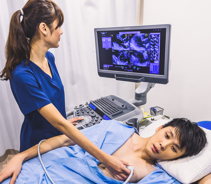
Echocardiography is a test that uses sound waves to produce live images of the heart. The image is called an echocardiogram. This test allows the doctor to monitor how the heart and its valves are functioning .
Why It’s done
The doctor may suggest an echocardiogram to;
- Check for problems with the valves or chambers of the heart
- Check if heart problems are the cause of symptoms such as shortness of breath or chest pain
- Detect congenital heart defects before birth (fetal echocardiogram)
Types of Echocardiogram
The type of echocardiogram a patient will have depends on the information the doctor needs.
Transthoracic Echocardiography
This is the most common type of echocardiography. It’s painless and noninvasive.
A device called a transducer will be placed on the patient’s chest over their heart. A computer interprets the sound waves as they bounce back to the transducer. This produces live images that are shown on a monitor
Transesophageal Echocardiography
If a transthoracic echocardiogram doesn’t produce definitive images or the patient needs to visualize the back of the heart better, the doctor may recommend a transesophageal echocardiogram.
The transducer tube is guided through the esophagus, the tube that connects the throat to the stomach. With the transducer behind the heart, the doctor can get a better view of any problems and visualize some chambers of the heart that are not seen on the transthoracic echocardiogram.
Stress Echocardiography
A stress echocardiogram uses traditional transthoracic echocardiography. However, the procedure is done before and after the patient has exercised or taken medication to make their heart beat faster. This allows the doctor to test how their heart performs under stress.
Three-dimensional Echocardiography
A three-dimensional (3-D) echocardiogram uses either transesophageal or transthoracic echocardiography to create a 3-D image of your heart. This involves multiple images from different angles. It’s used prior to heart valve surgery. It’s also used to diagnose heart problems in children.
Fetal Echocardiography
Fetal echocardiography is used on expectant mothers sometime during weeks 18 to 22 of pregnancy. The transducer is placed over the woman’s abdomen to check for heart problems in the fetus. The test is considered safe for an unborn child because it doesn’t use radiation, unlike an X-ray.
After the Procedure
Most people can resume their normal daily activities after an echocardiogram. If the echocardiogram is normal, no further testing may be needed.
If the results are concerning, the patient may be referred to a doctor trained in heart conditions (cardiologists) for more tests.
What We Offer
We at Almurshidi Medical Tourism will find the best doctors to cater to your needs. We are partnered with a wide network of hospitals and clinics that provide top quality medical experience.
We provide free medical estimates, make medical appointments, and provide several medical opinions if needed at no cost.
Contact Us
For more information contact us at +66822004040 or via WhatsApp


